Brain Plasticity Persists After Spinal Cord Injury During Rehab
THE PROTOHUMAN PERSPECTIVE#
This is the kind of finding that reframes what we think a damaged nervous system can do. For years, the implicit assumption in rehabilitation medicine has been a ceiling model: injury sets a hard limit, and therapy nudges you toward it. What Emmenegger and colleagues are showing is something different — that the ceiling might be partly illusory, at least at the structural level of cortical reorganization.
For anyone tracking human performance optimization, the implications extend well beyond spinal cord injury. If a brain undergoing active neurodegeneration can still mount plasticity responses equivalent to a healthy brain, what does that tell us about the untapped adaptive capacity in the rest of us? I think the word "rehabilitation" is doing too much work here. What we're really talking about is motor learning under extreme constraint — and the brain's refusal to stop adapting, even when the body's wiring is fundamentally disrupted. That matters whether you're recovering from a C5 injury or trying to build a new skill at fifty.
THE SCIENCE#
What Training-Induced Neuroplasticity Actually Looks Like After SCI#
Neuronal plasticity during motor rehabilitation is not a new concept. Draganski et al. demonstrated back in 2004 that training can induce grey matter changes in healthy adults[1]. But the question that lingered — and it's a genuinely important one — is whether this capacity survives severe injury. The nervous system after spinal cord injury isn't just idle. It's actively degenerating. Wallerian degeneration, demyelination, trans-synaptic atrophy — these processes continue for months and years post-injury[2].
Emmenegger et al. (2026) designed their study around this tension. Seventeen chronic SCI patients (all more than six months post-injury) and thirty-two healthy controls underwent training on a bimanual-bipedal computer-controlled device. The choice of a coordinated four-limb task is worth noting — it's not just moving one arm. It's demanding integrated sensorimotor processing across multiple systems simultaneously[3].
The central finding: SCI patients showed training-induced structural neuroplasticity that was comparable to, and in some regions greater than, healthy controls. This wasn't a subtle effect visible only in subgroup analysis. The injured brains adapted.
The Mechanism Question#
Here's where it gets complicated. What does "structural plasticity" actually mean at the tissue level? The study used quantitative MRI — specifically magnetization transfer saturation and other metrics sensitive to myelin and iron content — to track changes. These aren't functional activation maps showing which areas light up during a task. They're measuring actual tissue composition changes in grey and white matter.
I think this distinction matters more than most coverage will acknowledge. Functional MRI studies of SCI rehabilitation are common. They tell you the brain is doing something different. Structural MRI tells you the brain is becoming something different. The tissue itself is changing.
The fact that this occurs against a background of ongoing neurodegeneration suggests competing processes: degeneration pulling in one direction, training-driven plasticity pulling in another. The autophagy pathways that clear damaged cellular components, the myelination processes that rebuild signal transmission efficiency — these are active biological programs, not passive responses.
Converging Evidence: Cortical Reorganization Patterns#
Ma et al. (2025) published a case report that adds texture to this picture. A patient with incomplete cervical SCI underwent 20 sessions of robot-assisted bimanual training (RBMT). Before training, movement-related EEG desynchronization — a reliable index of sensorimotor cortex activation — was predominantly ipsilateral. After training, it shifted to contralateral dominance[4].
What does this actually feel like? The patient's brain was initially compensating by recruiting the wrong hemisphere. After training, it reorganized toward the normal pattern. That's not just a number on a screen — it represents a qualitative shift in how the brain processes movement intention.
But here's my skepticism: it's a single case. The effect is striking, and the EEG methodology is sound, but I'd want to see this replicated in a controlled cohort before treating it as definitive. The honest answer is that n=1 tells us what's possible, not what's typical.
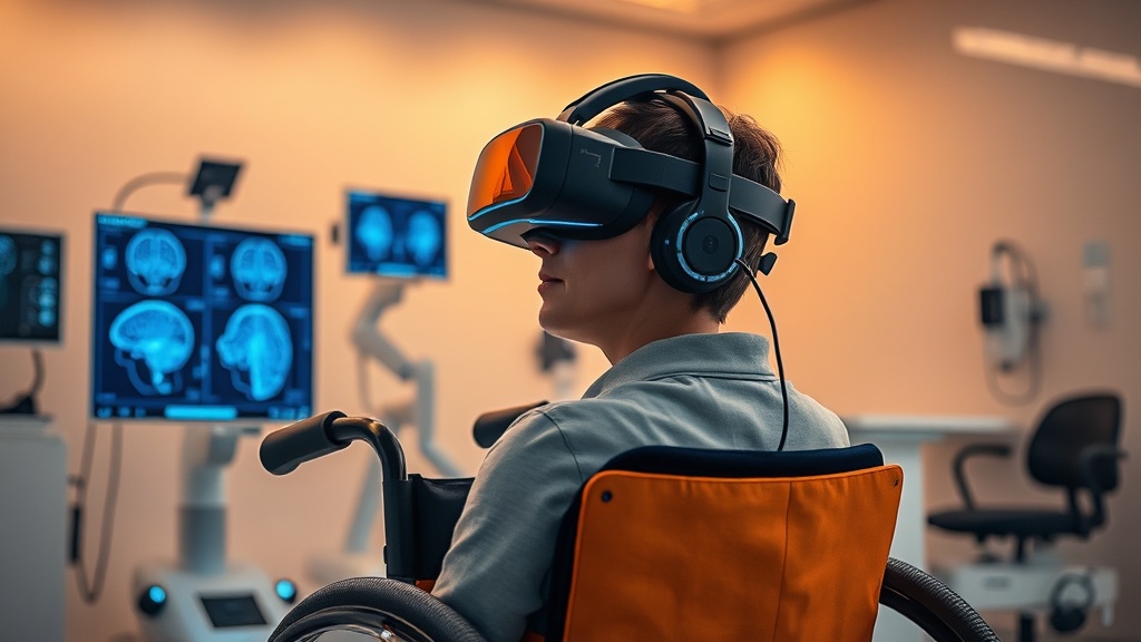
The VR-BCI Dimension#
Mannan et al. (2026) approached the problem differently, using virtual reality-mediated brain-computer interface training with both unimpaired individuals and SCI patients. Over multiple sessions, participants learned to modulate their sensorimotor EEG during motor imagery of virtual walking. BCI classification accuracy improved over time, indicating genuine learned neuromodulation[5].
The key contribution here is showing that even imagined movement — not physical rehabilitation — can drive measurable changes in sensorimotor processing after SCI. This matters enormously for patients with complete injuries who cannot perform physical movements at all.
Spinal Stimulation as an Amplifier#
Comino-Suárez et al. (2025) ran a proper double-blind, sham-controlled RCT — which is rarer than you'd think in this field — combining transcutaneous spinal cord stimulation (tSCS) with robotic-assisted gait training in 27 subacute incomplete SCI participants. The combination enhanced both motor scores and gait recovery compared to sham stimulation plus the same robotic training[6].
This is the most methodologically rigorous piece of the puzzle. The sham control is critical. Too many neuromodulation studies lack one, which makes it impossible to separate stimulation effects from placebo and natural recovery. Comino-Suárez et al. actually did the hard thing and still found a significant effect.
Rehabilitation Approaches and Evidence Quality in SCI Recovery
COMPARISON TABLE#
| Method | Mechanism | Evidence Level | Cost | Accessibility |
|---|---|---|---|---|
| Bimanual-bipedal training (Emmenegger) | Structural grey/white matter plasticity via coordinated motor learning | Moderate (n=49 controlled study) | Medium — specialized equipment required | Rehabilitation centers only |
| tSCS + robotic gait training (Comino-Suárez) | Spinal circuit neuromodulation combined with repetitive locomotor input | High (double-blind RCT, n=27) | High — robotic system + stimulation device | Specialized facilities |
| VR-mediated BCI training (Mannan) | Sensorimotor cortex modulation via motor imagery with visual biofeedback | Moderate (controlled, multi-session) | Medium — VR headset + EEG system | Expanding; consumer VR reducing cost |
| Robot-assisted bimanual training (Ma) | Cortical reorganization measured via ERD shift | Low (case report, n=1) | High — robotic rehabilitation device | Major rehabilitation hospitals |
| Standard physical therapy | Activity-dependent plasticity through repetitive movement | High (decades of RCTs) | Low–Medium | Widely available |
THE PROTOCOL#
Based on the current evidence — and I want to emphasize this is early-stage guidance, not settled clinical practice — here's how these findings translate into actionable steps for individuals pursuing neuroplasticity-driven rehabilitation after SCI.
Step 1: Establish baseline neurological assessment. Before beginning any structured rehabilitation protocol, obtain a comprehensive evaluation including ASIA Impairment Scale classification, motor scores for upper and lower extremities, and if possible, a baseline structural MRI. This isn't optional. Without knowing where you're starting, you cannot measure adaptation.
Step 2: Prioritize coordinated multi-limb training. The Emmenegger et al. data specifically used bimanual-bipedal tasks — not single-limb exercises. The coordinated demand appears to drive greater cortical engagement. Seek rehabilitation facilities with computer-controlled devices that allow synchronized upper and lower limb training, even if lower limb contribution is minimal or assisted.
Step 3: Consider transcutaneous spinal cord stimulation as an adjunct. Based on the Comino-Suárez RCT, tSCS applied at the lumbosacral level during robotic-assisted gait training enhanced outcomes. The stimulation parameters in their trial were non-invasive and well-tolerated. Discuss with your rehabilitation physician whether tSCS is appropriate for your injury level and completeness.
Step 4: Incorporate motor imagery with biofeedback. For sessions where physical training isn't possible — or for individuals with more complete injuries — VR-mediated BCI training using motor imagery of walking or limb movement may drive sensorimotor cortex modulation. The Mannan et al. protocol involved multiple sessions with real-time VR feedback congruent to imagined movements.
Step 5: Maintain training consistency over weeks, not days. The studies reviewed used protocols spanning multiple weeks — 20 sessions in the Ma case study, sustained multi-week programs in the Emmenegger and Comino-Suárez trials. Structural neuroplasticity requires sustained input. Sporadic training is unlikely to produce measurable tissue-level changes.
Step 6: Track progress with quantitative metrics. Where possible, incorporate objective outcome measures: motor scores, gait parameters (speed, step length, symmetry), and if accessible, follow-up neuroimaging. Subjective improvement matters, but the data here is built on quantifiable structural change.
Step 7: Address sleep and recovery as neuroplasticity enablers. While not the focus of the primary studies, neuroplasticity consolidation is sleep-dependent. Ensure adequate sleep quality — this is where the mitochondrial efficiency and NAD+ synthesis conversation becomes relevant, as cellular repair during sleep supports the structural remodeling these training protocols are driving.
Related Video
What is training-induced neuroplasticity in spinal cord injury?#
Training-induced neuroplasticity refers to structural and functional changes in brain tissue that occur in response to repeated, purposeful motor training. In SCI, this means the brain can physically reorganize — altering grey matter composition and cortical activation patterns — despite the fact that the spinal cord itself remains injured. The Emmenegger et al. study showed these changes are measurable with quantitative MRI and can match or exceed those seen in healthy individuals.
How does transcutaneous spinal cord stimulation enhance rehabilitation?#
Transcutaneous spinal cord stimulation delivers electrical current through the skin over the spine, targeting posterior spinal root nerve fibers. When combined with robotic-assisted gait training, tSCS appears to enhance the excitability of spinal circuits involved in locomotion, creating a more receptive neural environment for motor learning. The Comino-Suárez et al. RCT is currently the strongest evidence that this combination outperforms gait training alone.
Who benefits most from VR-mediated brain-computer interface training?#
Both unimpaired individuals and people with SCI showed improvements in sensorimotor neuromodulation during VR-BCI training in the Mannan et al. study. However, the approach may be particularly valuable for individuals with more severe or complete injuries who cannot perform physical movements — motor imagery doesn't require intact motor pathways to activate sensorimotor cortex. Optimal dosing and session frequency in humans are not yet firmly established.
Why does the injured brain sometimes show greater plasticity than healthy controls?#
This is one of the more surprising elements of the Emmenegger findings. One hypothesis is that the chronic SCI brain, having undergone deafferentation and cortical reorganization post-injury, exists in a state of heightened susceptibility to new input. It's somewhat analogous to how a sensory-deprived cortex may respond more dramatically when stimulation is reintroduced. But I'd be cautious — "greater" plasticity doesn't automatically mean "better" functional outcomes, and this relationship needs further investigation.
When should rehabilitation training begin after spinal cord injury?#
The Comino-Suárez trial focused on subacute patients, noting that this phase involves heightened neuroplastic potential that can be amplified by neuromodulation. However, the Emmenegger study worked with chronic patients (more than six months post-injury) and still found significant plasticity. The evidence suggests there isn't a hard window that closes — the nervous system retains adaptive capacity — but earlier intervention during the subacute phase may yield stronger effects due to greater endogenous neuroplastic activity.
VERDICT#
8/10. The Emmenegger et al. study is a genuinely important contribution — not because it tells us neuroplasticity exists (we knew that), but because it demonstrates that structural plasticity persists in a degenerating nervous system at levels comparable to healthy controls. That's a meaningful distinction. The converging evidence from tSCS trials, VR-BCI work, and bimanual training case studies strengthens the overall picture. I'm docking points because the sample sizes remain small (17 SCI patients in the lead study), the Emmenegger paper is still in unedited pre-publication form, and the direct link between structural MRI changes and functional recovery needs more work. The tSCS RCT from Comino-Suárez is the methodological standout. Overall, this is solid science with real clinical implications — but we're at the "promising and worth acting on cautiously" stage, not the "rewrite the textbooks" stage.
References
- 1.Draganski B, May A. Changes in grey matter induced by training. Nature (2004). ↩
- 2.Emmenegger TM, David G, Mohammadi S, Ziegler G, Callaghan MF, Thompson A, Friston KJ, Weiskopf N, Killeen T, Freund P. Longitudinal motor system changes from acute to chronic spinal cord injury. European Journal of Neurology (2024). ↩
- 3.Emmenegger TM, David G, Mohammadi S, Ziegler G, Callaghan MF, Thompson A, Friston KJ, Weiskopf N, Killeen T, Freund P. Neuronal plasticity during motor rehabilitation training after spinal cord injury. Communications Biology (2026). ↩
- 4.Ma D, Zeng RR, Chan SS, Pan Y, Zhang JJ. Case report: Movement-related neuroplasticity in a patient after spinal cord injury in response to task-oriented bimanual training. Frontiers in Human Neuroscience (2025). ↩
- 5.Mannan MMN, Palipana DB, Mulholland K, Jurd E, Lloyd ECR, Quinn ARJ, Crossley CB, Rabbi MF, Lloyd DG, Teng YD, Pizzolato C. Virtual reality mediated brain-computer interface training improves sensorimotor neuromodulation in unimpaired and post spinal cord injury individuals. Scientific Reports (2026). ↩
- 6.Comino-Suárez N, Moreno JC, Megía-García Á, del-Ama AJ, Serrano-Muñoz D, Avendaño-Coy J, Gil-Agudo Á, Gómez-Soriano J. Transcutaneous spinal cord stimulation combined with robotic-assisted body weight-supported treadmill training enhances motor score and gait recovery in incomplete spinal cord injury: a double-blind randomized controlled clinical trial. Journal of NeuroEngineering and Rehabilitation (2025). ↩
Fen Adler
Fen writes with psychological nuance and a slightly meandering quality that feels human. He'll start pursuing one idea, realize it connects to something else, and follow it briefly before returning: 'This reminds me of something from the attentional blink literature — different context, but the pattern holds.' He's interested in the experience, not just the mechanism, which means he'll occasionally ask: 'What does this actually feel like?' when discussing neurological effects.
View all articles →