Chronic Hypoxia Shrinks Hippocampus and Amygdala at Altitude
SNIPPET: Chronic hypoxia from long-term high-altitude living shrinks specific hippocampal and amygdalar subregions while preserving overall cortical volume, according to Chen et al. (2026) in Frontiers in Neuroscience. Despite measurable brain volume loss, migrants showed behavioral resilience — and longer residence paradoxically correlated with fewer insomnia symptoms, suggesting the brain adapts structurally under oxygen deprivation in ways we don't fully understand.
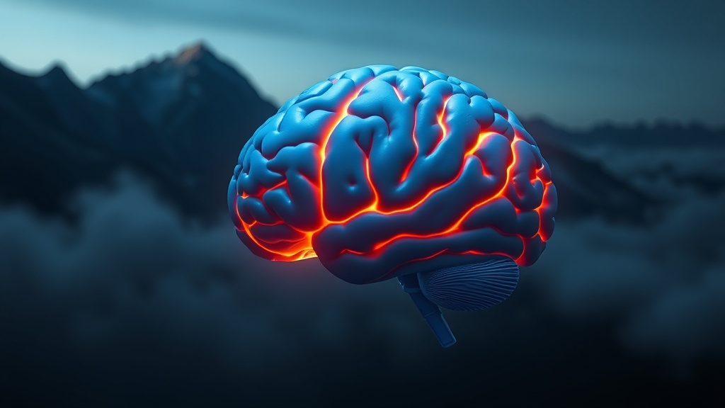
THE PROTOHUMAN PERSPECTIVE#
Your brain is the most oxygen-hungry organ you own. It accounts for roughly 2% of body mass and consumes 20% of your oxygen supply. Cut that supply — even mildly, even chronically — and the organ rewires itself. Not metaphorically. Physically.
What Chen et al. documented in 2026 is something that matters to anyone who trains at altitude, uses hypoxic tents, lives above 3,000 meters, or is considering intermittent hypoxia protocols for performance gains. The hippocampus and amygdala — your memory and threat-processing hardware — lose measurable volume under chronic low-oxygen conditions. But the cortex holds. And behavior, somehow, adapts.
This isn't a clean win-or-lose story. It's a trade-off story. And if you're manipulating your oxygen environment for performance, you need to understand what you're trading.
THE SCIENCE#
What Chronic Hypoxia Actually Does to Brain Structure#
Chronic hypoxia — sustained oxygen reduction over months or years — is not the same as the acute hypoxia you experience at the top of a hard interval session. It's a slow, persistent metabolic insult that forces the brain into adaptive compromises at the cellular level.
Chen et al. (2026) studied 44 young adult male Han Chinese migrants who relocated from low to high altitude, compared against 44 age-matched controls who remained at low altitude. Using high-resolution 3D structural MRI, the team mapped gray matter volume at both gross hemispheric and subregional levels within the hippocampus and amygdala [1].
The headline finding: bilateral cerebral cortex and cerebellar cortex volumes were essentially preserved in the high-altitude group. The brain's outer architecture held. But dig into the subcortical structures, and the damage is specific and targeted. Multiple hippocampal and amygdalar subregions showed significant volume reductions.
This is where it gets complicated.
Subregional Specificity — Not All Shrinkage Is Equal#
The hippocampus isn't a monolithic structure. It's a collection of subfields — CA1, CA3, the dentate gyrus, the subiculum — each with distinct functions in memory encoding, spatial navigation, and contextual fear processing. The amygdala has its own subregional architecture: the basolateral, centromedial, and superficial nuclei handle different emotional and threat-related computations.
Chen et al. found that the volume reductions weren't uniform across these subregions. Specific hippocampal and amygdalar subfields took the hit, while others were relatively spared [1]. This pattern suggests that chronic hypoxia doesn't just globally starve tissue — it selectively degrades regions with higher metabolic vulnerability, likely those with greater mitochondrial density and oxygen demand.
The mitochondrial efficiency story matters here. Hippocampal neurons, particularly in CA1 and the dentate gyrus, are among the most metabolically active cells in the brain. They depend on oxidative phosphorylation — aerobic ATP production — at rates that make them uniquely sensitive to oxygen deprivation. When NAD+ availability drops under hypoxic conditions, the electron transport chain falters, and these neurons are the first to suffer.

The Paradox: Behavioral Resilience#
Here's what I didn't expect from this data. Despite measurable structural losses, the high-altitude migrants showed what the authors call "behavioral resilience." Longer residence at high altitude was actually associated with fewer insomnia symptoms [1]. That's counterintuitive. You'd predict that more time under hypoxic stress would compound the cognitive and emotional damage. Instead, there's an adaptation curve.
I'm less convinced this means the brain is "fine." Volume loss in memory and emotion-processing regions doesn't just resolve because sleep improves. What it may indicate is that the brain compensates — rerouting functions, increasing synaptic efficiency in spared tissue, or activating autophagy pathways that clear damaged cells and allow surviving neurons to operate more cleanly. But these are hypotheses, not confirmed mechanisms.
The honest answer: we don't yet know why behavioral metrics improve while structural metrics decline. That gap is where the real science needs to happen.
Longitudinal Confirmation: The 4-Year Tibet Study#
This isn't an isolated finding. A separate 2026 longitudinal study tracked 69 college freshmen who moved from sea level to Tibet over four years. The results were less optimistic about behavioral resilience: persistent impairments in working memory and psychomotor function were documented throughout the exposure period [2].
Neuroimaging showed decreased gray matter volume and resting-state brain activity in the left putamen — a basal ganglia structure involved in motor control and procedural learning. Critically, the effect of exposure time on working memory was mediated by left putamen volume [2]. The structure shrank, and cognition declined in lockstep.
So we have two 2026 studies pointing at the same phenomenon — chronic hypoxia reduces subcortical gray matter — but diverging on the behavioral outcome. Chen et al. found resilience; the Tibet longitudinal study found persistent deficits. The difference may come down to the specific regions affected, the altitude and duration of exposure, or individual variation in hypoxic adaptation.
The Mechanistic Framework: Inflammation, BBB, and Neuroplasticity#
A comprehensive 2026 review in the Journal of Neuroinflammation helps contextualize these findings. The authors describe two main pathways through which chronic mild hypoxia affects the brain: direct effects on neural tissue and systemic inflammation that compromises the blood-brain barrier (BBB) [3].
Chronic hypoxia degrades BBB integrity, allowing peripheral inflammatory molecules into brain tissue that's normally protected. This creates a neuroinflammatory environment that accelerates neuronal damage — particularly in regions already stressed by metabolic demand. At the same time, mild hypoxia can activate adaptive responses: HIF-1α stabilization drives angiogenesis, erythropoietin production, and shifts in metabolic substrate utilization that may temporarily protect tissue [3].
The dual nature of this response — damage and adaptation running simultaneously — is what makes hypoxia protocols so tricky to prescribe.
Vagus Nerve Stimulation: A Countermeasure?#
One of the more promising interventions comes from Sharma et al. (2025), who tested vagus nerve stimulation (VNS) in rats exposed to severe hypoxia (8% oxygen). VNS during hypoxia exposure restored NGF mRNA to baseline levels in the hippocampus and partially recovered BDNF mRNA [5]. On behavioral testing, VNS-treated animals showed significantly less cognitive impairment than hypoxia-only animals.
Let me push back on this slightly: this is a rat model using acute severe hypoxia, not chronic mild hypoxia in humans. The translation gap is real. But the mechanism — VNS upregulating neurotrophic factors in the hippocampus — is biologically plausible and worth tracking.
Brain Regions Affected by Chronic High-Altitude Hypoxia
COMPARISON TABLE#
| Method | Mechanism | Evidence Level | Cost | Accessibility |
|---|---|---|---|---|
| Long-term altitude residence | Chronic hypoxic adaptation; subregional gray matter remodeling | Two 2026 human neuroimaging studies (n=88, n=69) | Free (geographic) | Limited to high-altitude locations |
| Intermittent hypoxic training (IHT) | Controlled oxygen cycling; HIF-1α activation | Multiple small human trials; mixed results | $200–$2,000 (masks/tents) | Moderate — requires equipment |
| Altitude simulation masks | Restricted airflow; mild inspiratory resistance | Weak evidence; minimal true hypoxic stimulus | $30–$150 | High — consumer available |
| Vagus nerve stimulation (VNS) | Neurotrophin upregulation (NGF, BDNF) in hippocampus | Preclinical rat model (2025) | $100–$500 (transcutaneous devices) | Moderate — consumer devices exist |
| Aerobic exercise at sea level | Increased cerebral perfusion; BDNF upregulation; angiogenesis | Strong — multiple RCTs and meta-analyses | Free–low | Universal |
THE PROTOCOL#
Protecting Brain Structure Under Hypoxic Exposure: An Evidence-Based Approach
If you live, train, or work at altitude — or if you're using hypoxic protocols for performance — these steps are based on current evidence for mitigating subcortical volume loss.
Step 1: Establish your baseline. Before any extended altitude exposure or hypoxic training protocol, get a cognitive baseline. Use validated tools: the Montreal Cognitive Assessment (MoCA) for general screening, and a working memory task like the N-back. Record your HRV baseline. You can't track decline without a reference point.
Step 2: Monitor sleep architecture aggressively. Chen et al. found that insomnia severity correlated with hippocampal and amygdalar subregional volume changes [1]. Track sleep with a device that measures HRV and sleep staging — not just duration. If insomnia symptoms increase in the first 8–12 weeks of altitude exposure, intervene early.
Step 3: Prioritize aerobic exercise at altitude. This is non-negotiable. Aerobic training at altitude increases cerebral blood flow, upregulates BDNF, and supports angiogenesis — all of which counteract the mechanisms driving subcortical volume loss. Aim for 150–200 minutes per week of zone 2 cardio, adjusted for altitude-related performance reduction.
Step 4: Consider transcutaneous vagus nerve stimulation (tVNS). Based on the Sharma et al. (2025) preclinical data showing hippocampal neurotrophin restoration with VNS [5], tVNS devices (auricular stimulation) may offer a low-risk adjunct. Use 25–30 Hz stimulation, 15–20 minutes daily. This is early-stage evidence — I'd want to see human replication before calling it essential, but the risk profile is minimal.

Step 5: Supplement for mitochondrial support. Under chronic hypoxia, mitochondrial efficiency drops. NAD+ precursors (NMN at 500 mg/day or NR at 300 mg/day) may support the electron transport chain under oxygen-limited conditions. Add CoQ10 (200 mg/day) and magnesium glycinate (400 mg before sleep) to support both mitochondrial function and sleep quality. These are based on general mitochondrial support evidence — not altitude-specific trials.
Step 6: Cycle your hypoxic exposure if it's voluntary. If you're using altitude training or hypoxic tents by choice, don't live in them continuously. The data from Chen et al. shows a clear relationship between duration of high-altitude exposure and subregional volume loss [1]. Build in normoxic recovery periods — at least 2 weeks at sea level for every 8–10 weeks of sustained altitude exposure.
Step 7: Re-test cognition every 3 months. Compare against your baseline. If working memory scores drop more than 15% or psychomotor speed declines measurably, reduce hypoxic exposure and increase the interventions above.
Related Video
What brain regions are most vulnerable to chronic hypoxia?#
The hippocampus and amygdala — specifically their subregions — appear most vulnerable based on Chen et al. (2026) and the Tibet longitudinal study. These structures have high metabolic demands and rely heavily on oxidative phosphorylation. The cerebral cortex, surprisingly, maintains its volume even under prolonged hypoxic exposure, suggesting differential metabolic resilience across brain regions [1][2].
How long does it take for altitude-related brain changes to appear?#
The 4-year Tibet study documented progressive gray matter reductions in the left putamen across years two and four of high-altitude residence [2]. Chen et al. also found that duration of high-altitude exposure correlated with subregional volume changes. Meaningful structural changes likely emerge within the first 1–2 years, though individual variation is significant.
Can the brain recover after returning to low altitude?#
Honestly, we don't have clean data on this yet. The studies focus on ongoing exposure, not recovery trajectories. Based on what we know about neuroplasticity and gray matter remodeling, partial recovery is plausible — particularly if the individual is young and engages in interventions that support neurogenesis (exercise, sleep optimization). But I'd want to see longitudinal return-to-sea-level imaging before making promises.
Why did longer altitude residence correlate with better sleep?#
This is one of the more puzzling findings from Chen et al. It may reflect acclimatization — the body adapts its ventilatory response, erythropoietin production, and hemoglobin levels over time, improving oxygen delivery during sleep. It could also reflect a survivor bias: those who tolerate altitude best may remain longest. The mechanism isn't established [1].
Does vagus nerve stimulation work for hypoxia-related cognitive decline in humans?#
Not yet proven in humans. Sharma et al. (2025) showed VNS restored hippocampal NGF expression and reduced cognitive impairment in rats exposed to acute severe hypoxia [5]. The mechanism is promising — neurotrophin upregulation in precisely the structures that chronic hypoxia damages. But translating from acute rat hypoxia at 8% oxygen to chronic human altitude exposure requires significant further research.
VERDICT#
Score: 7/10
The Chen et al. (2026) study delivers something genuinely new: subregional mapping of hippocampal and amygdalar volume loss under chronic altitude exposure, paired with the unexpected behavioral resilience finding. The specificity of the structural data is valuable. Combined with the Tibet longitudinal study and the neuroinflammation review, we now have a converging picture of how chronic hypoxia reshapes subcortical brain architecture.
But the sample is small (n=44 per group), male-only, and cross-sectional — which means we're looking at a snapshot, not a trajectory. The behavioral resilience claim is interesting but underexplained mechanistically. And the protocol implications are still more theoretical than proven.
I take this seriously because I've spent time at altitude and felt the cognitive fog that doesn't show up on a standard wellness check. The data here validates what a lot of high-altitude workers and athletes already suspect: something is happening to the brain up there, and it's not just acute mountain sickness. It's structural. Whether it's reversible — that's the question worth funding.
References
- 1.Chen X, Huang J, Li H, Li G, Zhu J, Du S, Zhou J, Du F. Chronic hypoxia reshapes the brain: subregional volume reductions of the hippocampus and amygdala contrasts with behavioral resilience in long-term high-altitude migrants. Frontiers in Neuroscience (2026). ↩
- 2.Author(s) not listed. Chronic high-altitude exposure and cognitive health in Chinese college students: a 4-year longitudinal neuroimaging study. Scientific Reports (2026). ↩
- 3.Author(s) not listed. Mild chronic hypoxia and the brain: an ambiguous relationship. Journal of Neuroinflammation (2026). ↩
- 4.Jiao D. From hypoxic pockets to daily routines: linking brain oxygenation and cognitive resilience. Frontiers in Aging Neuroscience (2025). ↩
- 5.Sharma B, Jones KA, Olsen LK, Moore RJ, Curtner FS, Hatcher-Solis CN. Vagus nerve stimulation ameliorates cognitive impairment caused by hypoxia. Frontiers in Behavioral Neuroscience (2025). ↩
Cira Renn
Cira writes with physical conviction — she's done this, she knows what it feels like, and she doesn't pretend otherwise. Her writing has visceral energy: 'Cold water at 10°C isn't a wellness trend. It's a physical confrontation.' She distinguishes between what the research shows and what she's experienced, and she'll tell you when they diverge.
View all articles →