Photobiomodulation Boosts Meniscus Stem Cell Growth via TRPV1
SNIPPET: Photobiomodulation (PBM) at 700–710 nm and 1064 nm wavelengths with 15 J/cm² energy density significantly enhances mitochondrial function and proliferation in human meniscus-derived stem cells (MeSCs) through TRPV1 calcium channel activation. Higher energy densities and shorter wavelengths reduced cell viability, confirming PBM's narrow therapeutic window for regenerative applications.
THE PROTOHUMAN PERSPECTIVE#
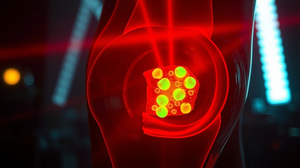
Meniscus injuries wreck knees. They wreck careers, mobility, independence. Surgery success rates remain frustratingly low, and the tissue's natural self-repair capacity is almost nonexistent. So when a study maps the precise wavelength-and-dose parameters that drive stem cell proliferation in meniscus tissue — through a defined molecular pathway, not hand-waving — I pay attention.
This matters for the optimization crowd because it represents the intersection of two things we care about: light therapy done with actual precision, and stem cell biology moving beyond the hype cycle. The data here doesn't just show that PBM "works" — it shows exactly where the dose-response curve collapses. Most conditions tested actually harmed the cells. That's the kind of specificity the consumer PBM market pretends doesn't exist.
Combined with emerging research on telomere dynamics in mesenchymal stromal cells and the longevity potential of MSC-derived extracellular vesicles, we're looking at a converging picture: the tools for tissue regeneration are sharpening, but only if you respect the parameters.
THE SCIENCE#
Photobiomodulation and Meniscus Stem Cells: The TRPV1 Connection#
Photobiomodulation is the application of specific light wavelengths to modulate cellular behavior — not heat therapy, not generic "red light." The distinction matters. Published in Scientific Reports (Nature) in December 2025, the study by researchers investigating human meniscus-derived stem cells (MeSCs) tested four wavelength bands (400–405, 500–505, 700–710, and 1064 nm) across four energy densities (3, 15, 30, and 60 J/cm²) [1].
The results were unambiguous in one respect: only the 700–710 nm and 1064 nm wavelengths at energy densities of 3, 15, and 30 J/cm² improved MeSC proliferation, with 15 J/cm² producing the most significant effect. Every other combination — shorter wavelengths, higher energy densities — actually reduced mitochondrial function and proliferative capacity.
Let me be blunt about what this means. If you're pointing a 405 nm LED at your knee and expecting cartilage regeneration, you're likely doing more harm than good at the cellular level.
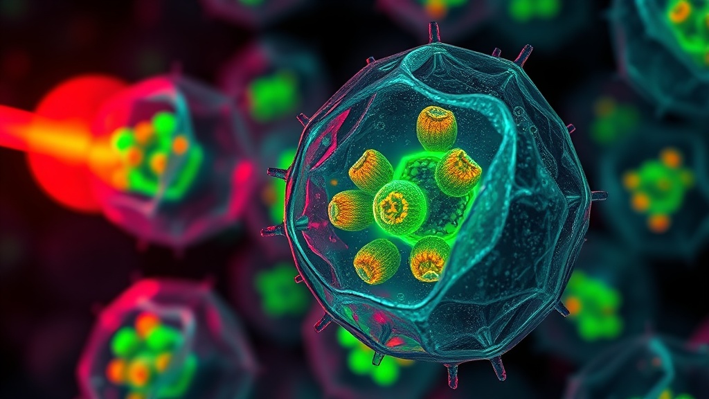
The Molecular Mechanism: Calcium, ROS, and the Biphasic Response#
The researchers didn't stop at observing proliferation changes. They mapped the signaling cascade. Intracellular calcium (Ca²⁺) levels and reactive oxygen species (ROS) production both increased with energy density across all wavelengths. But here's where it gets interesting — cytochrome C oxidase (CCO) activity and nitric oxide (NO) concentrations remained unchanged [1].
This challenges the dominant narrative in PBM literature. The prevailing model holds that PBM works primarily through photodissociation of NO from CCO in the mitochondrial electron transport chain. These data suggest a different primary mechanism: TRPV1 calcium channel activation.
When the researchers inhibited TRPV1 channels pharmacologically, the PBM-induced elevations in Ca²⁺ and ROS were abolished at all wavelengths. Proliferation changes — both positive and negative — were prevented. The entire dose-response curve collapsed to baseline [1].
This is a TRPV1-Ca²⁺-ROS signaling axis. Not CCO. Not NO dissociation. The implications for protocol design are significant.
The Biphasic Dose Problem#
This study is a textbook demonstration of hormesis — the biphasic dose response where low doses stimulate and high doses inhibit. At 60 J/cm², even the favorable wavelengths produced excessive ROS that overwhelmed cellular antioxidant capacity and collapsed mitochondrial membrane potential. The therapeutic window isn't just narrow; it's a cliff edge.
I'm less convinced by one aspect of the study design: the energy densities tested jump from 30 to 60 J/cm² with nothing in between. The optimal dose might sit at 20 or 25 J/cm², but we simply don't have that resolution. Worth noting.
Telomere Dynamics and Cell Size Selection#
Supporting the broader regenerative picture, Berzins et al. (2026) published data in Frontiers in Bioengineering and Biotechnology demonstrating that smaller adipose-derived mesenchymal stromal cells (AD-MSCs) possess significantly longer telomeres — 18,121 base pairs versus 15,870 bp in the general population — and express pluripotency markers Nanog and OCT3/4 [2].
A 14.2% increase in telomere length from size selection alone. These smaller cells also formed teratoma-like structures with skin-like morphology in immunodeficient mice, suggesting genuine pluripotent capacity. The practical implication: cell size could serve as a non-destructive proxy for telomere length, enabling real-time quality control in regenerative protocols without sacrificing cells for measurement.
MSCs as Longevity Tools: The Extracellular Vesicle Angle#
Rudnitsky, Braiman, Wolfson et al. published a review in Biogerontology (2025) synthesizing the evidence for MSCs and their derivatives — particularly extracellular vesicles (EVs) — as longevity-promoting tools [3]. The standout finding: over 70% of microRNAs over-represented in embryonic stem cell-derived EVs target longevity-associated genes (LAGs). Systemic administration of MSCs and EVs modified the omic profiles of aged rodent organs toward younger signatures.
The catch, though. MSC efficacy appears relatively independent of donor age in humans but not in rodents. This species discrepancy should give us pause before extrapolating too aggressively from mouse lifespan studies.
Scalable Secretome Production: The Hair Follicle Platform#
A November 2025 study in Stem Cell Research & Therapy developed immortalized human hair follicle-derived mesenchymal-like stromal cells (iHF-MSCs) that produce consistent therapeutic secretome enriched in immunoregulatory factors (Gal-9, IL-1Ra, TSG-6) and pro-regenerative factors (VEGF, PDGF-AA, EGF) [4]. From 576 single-cell clones, two (C18 and C26) were selected for long-term stability and preserved multipotency.
This addresses the perennial manufacturing bottleneck in MSC therapy: donor variability and replicative senescence. Cell-free secretome therapies bypass many regulatory hurdles that direct cell therapy faces.
MeSC Proliferation Response by PBM Wavelength at 15 J/cm²
COMPARISON TABLE#
| Method | Mechanism | Evidence Level | Cost | Accessibility |
|---|---|---|---|---|
| PBM at 700-710 nm / 15 J/cm² | TRPV1-Ca²⁺-ROS signaling → MeSC proliferation | Single in vitro study (Nature SR) | Low–Moderate (clinical LED devices) | Clinic-based; some consumer devices approximate parameters |
| PBM at 660 nm (standard red) | CCO photodissociation / NO release (traditional model) | Multiple in vitro and small human trials | Low (consumer panels widely available) | High — widely available consumer devices |
| MSC injection therapy | Direct cell transplantation, paracrine signaling | Multiple Phase I/II trials; mixed results | High ($5,000–$25,000+ per treatment) | Limited — specialized clinics only |
| MSC-derived EVs / secretome | Cell-free paracrine factors, miRNA cargo | Preclinical + early clinical | Moderate–High (manufacturing dependent) | Very limited — mostly research stage |
| Size-selected AD-MSCs (longer telomeres) | Enhanced replicative capacity, pluripotency marker expression | Single preclinical study | High (specialized isolation) | Research only |
| iHF-MSC secretome platform | Immortalized cell line → scalable secretome production | Single preclinical study | Potentially moderate at scale | Research only |
THE PROTOCOL#
Based on the current evidence — and I want to emphasize this is derived from a single in vitro study for the PBM parameters — here is a framework for those tracking this space or working with clinicians on knee regeneration.
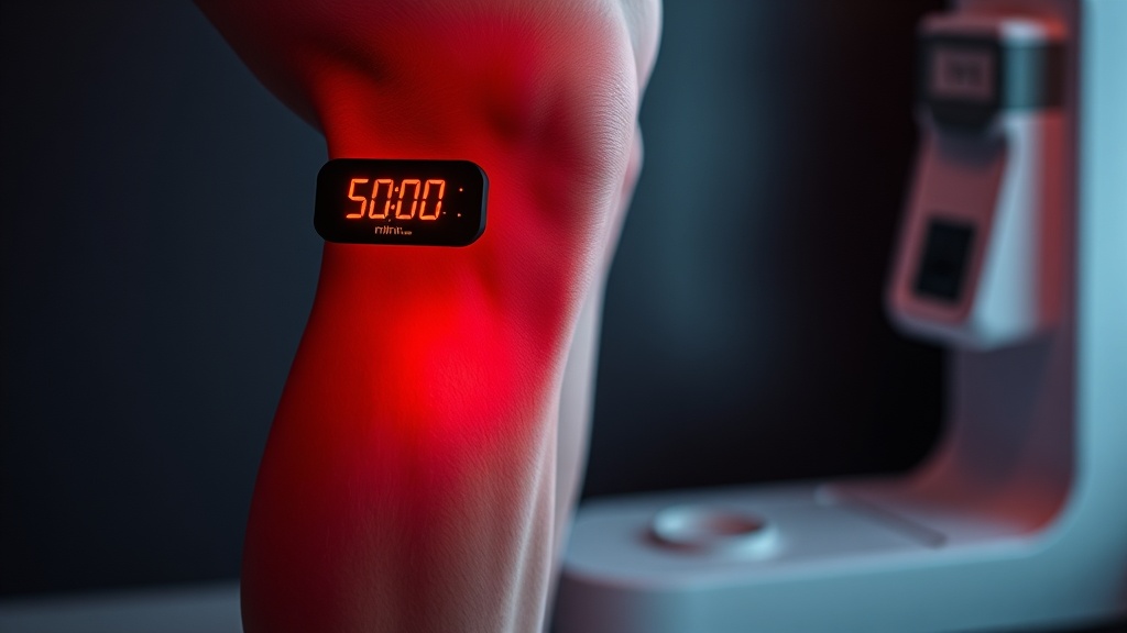
Step 1. Confirm your light source specifications before anything else. You need a device capable of delivering 700–710 nm or 1064 nm wavelengths. Not "approximately red." Not broadband. Check the manufacturer's spectral output data. Most consumer panels operate at 630–660 nm, which is not the wavelength range that showed benefit in this study.
Step 2. Calculate your energy density delivery. Target 15 J/cm² at the tissue surface. This requires knowing your device's irradiance (mW/cm²) and applying the formula: Energy Density (J/cm²) = Irradiance (W/cm²) × Time (seconds). For a device delivering 50 mW/cm², you'd need 300 seconds (5 minutes) of exposure. Exceeding 30 J/cm² reversed the benefits entirely.
Step 3. Position the light source with minimal distance to the knee joint. Tissue penetration drops exponentially with depth and is further reduced by skin pigmentation, subcutaneous fat, and clothing. Direct skin contact with a calibrated device is the baseline assumption in this research.
Step 4. Frequency of application remains unestablished. The in vitro study used single-exposure protocols. For anyone choosing to experiment, I'd suggest starting with every 48–72 hours and monitoring subjective joint comfort — but honestly, optimal human dosing frequency is not yet known for this specific application.
Step 5. If pursuing MSC-based regenerative approaches in parallel, discuss size-selection protocols with your provider. The Berzins et al. data suggests smaller AD-MSCs carry longer telomeres (18,121 bp vs. 15,870 bp) and enhanced regenerative markers [2]. This is not yet standard practice, but the selection method is straightforward.
Step 6. Track outcomes objectively. MRI-based cartilage assessment, validated knee function scores (KOOS, Lysholm), and ideally, biomarkers of cartilage turnover (CTX-II, COMP) provide meaningful endpoints. Subjective pain reduction alone is insufficient to evaluate tissue-level changes.
Related Video
What wavelength of light therapy works best for meniscus stem cell proliferation?#
Based on the 2025 Scientific Reports study, 700–710 nm and 1064 nm wavelengths at 15 J/cm² energy density produced the strongest proliferative response in human meniscus-derived stem cells. Shorter wavelengths (400–505 nm) and energy densities above 30 J/cm² actually impaired cell viability and mitochondrial function. Most consumer red light devices operate at 630–660 nm, which was not tested in this specific study.
How does TRPV1 channel activation relate to photobiomodulation?#
TRPV1 (transient receptor potential vanilloid 1) is a calcium channel that, in this study, served as the primary mediator of PBM's effects on meniscus stem cells. When researchers blocked TRPV1 pharmacologically, PBM-induced increases in intracellular calcium and ROS were eliminated, and all proliferative changes were prevented [1]. This suggests TRPV1-Ca²⁺-ROS signaling, rather than the traditional cytochrome C oxidase pathway, drives PBM's effects in these cells.
Why do smaller mesenchymal stem cells have longer telomeres?#
Berzins et al. found that smaller AD-MSCs had telomeres averaging 18,121 bp compared to 15,870 bp in the total population, along with expression of pluripotency markers Nanog and OCT3/4 [2]. The likely explanation is that smaller cells represent a less differentiated, more primitive subpopulation with greater replicative reserve. Cell size may function as a practical, non-destructive biomarker for selecting higher-quality cells in regenerative protocols.
What are MSC-derived extracellular vesicles and can they extend lifespan?#
Extracellular vesicles (EVs) are nanoscale particles secreted by cells, carrying proteins, lipids, and microRNAs that modulate recipient cell behavior. Rudnitsky et al.'s review found that over 70% of microRNAs enriched in ESC-derived EVs target longevity-associated genes, and systemic EV administration shifted aged rodent tissue profiles toward younger signatures [3]. However, this evidence is entirely preclinical — human lifespan extension data does not yet exist.
When will PBM-based meniscus regeneration be available clinically?#
The honest answer: not soon. The MeSC-PBM data is in vitro — cultured cells in a lab, not living knee joints with intact tissue architecture, blood supply, and immune responses. Clinical translation requires animal model validation, safety profiling, and human trials. I'd estimate 5–10 years before any protocol based specifically on these findings reaches clinical practice, though off-label PBM use for joint conditions already exists in some clinics.
VERDICT#
Score: 7/10
The PBM-MeSC study is genuinely useful because it identifies a specific molecular mechanism — TRPV1-Ca²⁺-ROS — and maps a clear dose-response curve with both beneficial and harmful parameter zones. That's more than most PBM papers deliver. The supporting MSC research on telomere dynamics and secretome platforms adds context to where regenerative medicine is heading.
But this is a single in vitro study. No animal models, no human data, no long-term follow-up. The energy density resolution between 30 and 60 J/cm² is too coarse. And the disconnect from the traditional CCO-NO model needs replication before I'd rewrite my understanding of PBM mechanisms entirely. The foundations here are solid — the clinical applications remain speculative.
References
- 1.Author(s) not listed. Photobiomodulation stimulates mitochondrial function and cell proliferation in meniscus-derived stem cells (MeSCs) via activation of TRPV1 channel. Scientific Reports (2025). ↩
- 2.Berzins U, Shishkin A, Zvirgzdina R, Rautmane A, Baronins J, Sorokins H, Goel S, Minchev D. Small adipose-derived mesenchymal stromal cells exhibit longer telomeres and enhanced regenerative potential. Frontiers in Bioengineering and Biotechnology (2026). ↩
- 3.Rudnitsky E, Braiman A, Wolfson M, Muradian KK, Gorbunova V, Turgeman G, Fraifeld VE. Mesenchymal stem cells and their derivatives as potential longevity-promoting tools. Biogerontology (2025). ↩
- 4.Author(s) not listed. Immortalized human hair follicle-derived mesenchymal-like stromal cells for the long-term production of scalable immunomodulatory and regenerative secretome. Stem Cell Research & Therapy (2025). ↩
Sova Reld
Sova writes with focused intensity and low tolerance for vague claims. She came to photobiomodulation through personal experimentation and is irritated by both true believers and reflexive skeptics. Her writing has edge: 'The wellness market has done more damage to this field than the skeptics ever could.' She's extremely precise about parameters — wavelength, irradiance, duration — and will tell you when a study used inadequate dosing without apology.
View all articles →