Janus Particles Detect Cancer Vesicles in Under 60 Minutes
SNIPPET: Janus particles with fluorescent half-shells detect cancer-derived small extracellular vesicles in under 60 minutes from less than 10 μl of unprocessed plasma, achieving AUC values of 0.90–0.99 across colorectal cancer, pancreatic adenocarcinoma, glioblastoma, and Alzheimer's disease — with 100× better sensitivity than ultracentrifugation-based methods.
THE PROTOHUMAN PERSPECTIVE#
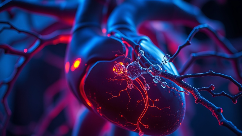
Liquid biopsy has been the white whale of early cancer detection for over a decade. The premise is simple: cancer cells shed molecular debris into your bloodstream long before a tumor becomes visible on imaging. Catch those signals early enough, and you catch the disease before it catches you.
The problem has always been the catching part. Small extracellular vesicles — tiny membrane-bound packets released by cells — carry disease-specific surface markers, but they're absurdly difficult to isolate from the protein noise in blood. Current gold-standard methods require ultracentrifugation, 24+ hours of processing, and expensive instrumentation. That's a research tool, not a clinical one.
This new Janus particle assay collapses that workflow into 60 minutes with a finger-prick volume of sample. For anyone tracking their biomarkers or pushing for earlier disease screening, this is the kind of platform shift that turns liquid biopsy from a concept into something your doctor might actually order. The distinction matters: a test that exists in a lab is interesting. A test that works in a clinic is actionable.
THE SCIENCE#
What Are Small Extracellular Vesicles, and Why Should You Care?#
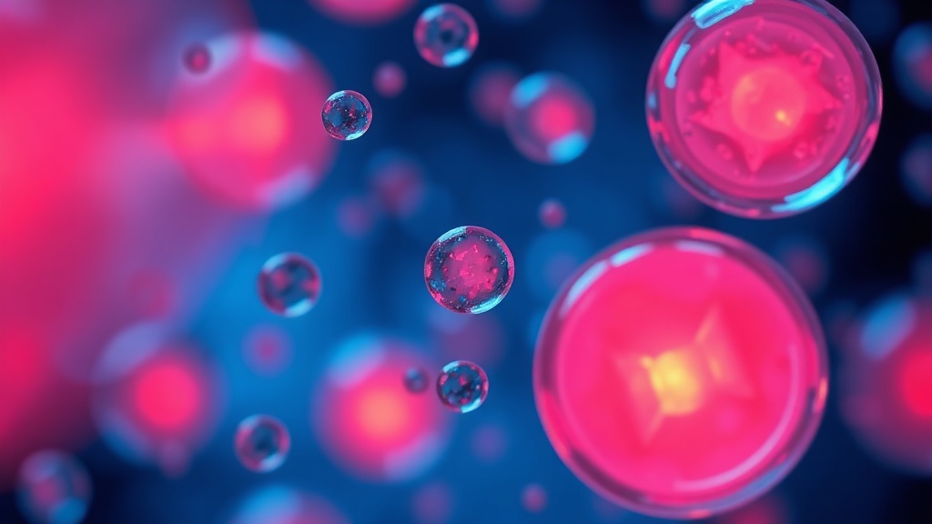
Small extracellular vesicles (sEVs), including exosomes, are nanoscale membrane particles (30–150 nm) released by virtually all cells in the body. They carry surface proteins and molecular cargo that reflect their cell of origin — which means a vesicle shed by a pancreatic tumor cell looks different from one released by a healthy hepatocyte[1]. This makes them, in theory, ideal biomarkers for disease detection. In practice, isolating them from blood has been a nightmare.
Conventional approaches — western blot, ELISA, bead-based flow cytometry — require large sample volumes, extensive purification steps, and cannot assess individual vesicle heterogeneity[3]. Ultracentrifugation combined with surface plasmon resonance (UC + SPR) is sensitive but takes roughly 24 hours and demands significant sample processing. None of this scales to a clinical screening context.
The Janus Particle Trick#
Here's where it gets clever — and I'll admit, the physics of this one is elegant.
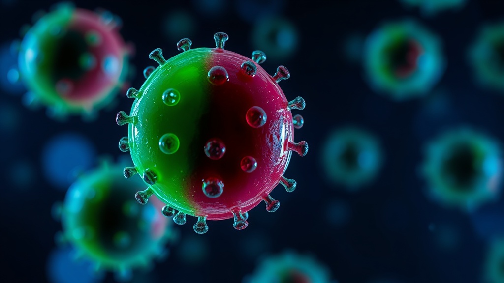
Janus particles are micrometre-sized spheres with two distinct hemispheres: one fluorescent, one dark. In solution, they undergo Brownian rotation, producing a characteristic "blinking" pattern as the fluorescent side rotates in and out of the detection plane. The key insight from this Nature Biomedical Engineering study is that when a small extracellular vesicle binds to the particle surface, the altered hydrodynamic drag changes the blinking frequency in a measurable way[1].
Smaller proteins — the abundant background noise in plasma — don't produce a detectable change. Their mass is too low to shift the rotational dynamics. This gives the assay an inherent size-selectivity without requiring any isolation or purification step, which is annoying, actually, because it makes you wonder why nobody exploited this physics earlier.
The assay uses immunological Janus particles (IJPs) functionalized with antibodies against sEV surface markers. When sEVs bind, the blinking rate shifts. The magnitude of that shift correlates with vesicle concentration and surface marker density[1].
The Numbers That Matter#
Let me be specific about what this assay delivers, because the performance gap over existing methods is striking.
Detection limit: approximately 200 vesicles per microlitre. That's two orders of magnitude more sensitive than UC + SPR[1].
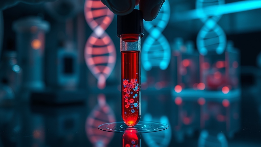
Sample volume: less than 10 μl. For context, a standard blood draw is 5–10 ml. This requires a thousandth of that.
Time to result: under 60 minutes, versus 24 hours for UC + SPR.
Sample compatibility: works directly on plasma, serum, urine, and cell culture media — no pre-processing required.
In the blind clinical study, the assay was tested on 87 human subjects across five groups: colorectal cancer (CRC), pancreatic ductal adenocarcinoma (PDAC), glioblastoma (GBM), Alzheimer's disease (AD), and healthy controls. The method identified disease type with AUC values ranging from 0.90 to 0.99[1]. An AUC of 0.99 is, frankly, exceptional for a liquid biopsy platform at this stage of development.
But here's where I want to push back slightly. Eighty-seven subjects is not a large cohort. It's a proof-of-concept study — a strong one — but the jump from 87 to 8,700 subjects has historically been where many promising biomarker assays stumble. The AUC numbers are impressive, but I'd want to see these replicated in a multi-center trial before calling this clinically validated. The honest answer is we don't know how this performs in populations with comorbidities, early-stage cancers, or confounding inflammatory conditions.
The Brownian Rotation Mechanism in Detail#
The physics underpinning this assay relies on the relationship between particle size, surface drag, and rotational diffusion. Brownian motion is stochastic, but the rotational diffusion coefficient for a sphere is inversely proportional to the cube of its effective radius. When sEVs (averaging ~100 nm) bind to a micrometre-scale Janus particle, they increase the effective hydrodynamic radius enough to produce a measurable decrease in blinking frequency.
The transport-limited nature of binding means the assay responds to actual vesicle concentration in solution rather than requiring equilibrium binding — a practical advantage for rapid measurement[1]. Wang et al. demonstrated a complementary approach using magnetic Janus particles with active electromagnetic driving, where a cutoff frequency shift served as the detection signal, achieving sEV concentrations of 2.9 × 10⁹ particles/mL with the CD63 surface marker[2].
Janus Particle Assay vs. UC + SPR: Performance Comparison
COMPARISON TABLE#
| Method | Mechanism | Evidence Level | Cost | Accessibility |
|---|---|---|---|---|
| Janus Particle (IJP) Assay | Brownian rotation blinking frequency shift upon sEV binding | Single blind study, 87 subjects, AUC 0.90–0.99 | Estimated low (minimal equipment) | Research stage; not yet commercially available |
| UC + SPR | Ultracentrifugation isolation + surface plasmon resonance | Established research method, widely validated | High (expensive instrumentation) | Specialized labs only |
| PANORAMA Microfluidic | Plasmonic nano-aperture label-free imaging on gold nanodisks | Single study, 20 μL plasma, 60 min | Moderate (custom microfluidic chip) | Research stage |
| Magnetic Janus Particles (MJP) | Electromagnetic cutoff frequency shift upon sEV binding | Single study, CD63 marker validated | Low-moderate | Research stage |
| Bead-based Flow Cytometry | Fluorescence-labeled bead capture and sorting | Widely validated, limited single-vesicle resolution | Moderate-high | Clinical and research labs |
| ELISA for sEV Markers | Antibody sandwich immunoassay | Well-established but bulk measurement only | Low-moderate | Widely accessible |
THE PROTOCOL#
This section is framed for researchers, clinicians, and advanced biohackers tracking the liquid biopsy space. The IJP assay is not yet commercially available, but based on the published methodology, here is how the workflow operates — and how to prepare for its eventual clinical translation.
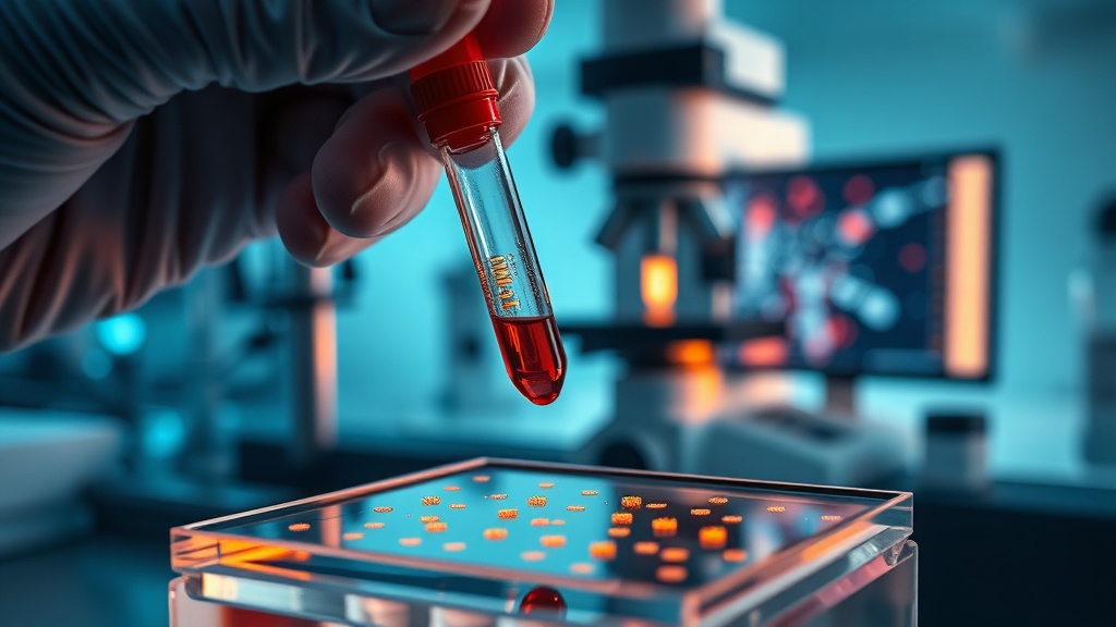
Step 1: Sample Collection Collect less than 10 μl of plasma, serum, or urine. No pre-processing, centrifugation, or purification is required. Standard phlebotomy or finger-prick blood collection is sufficient for plasma samples. Store samples according to standard biobanking protocols if not processing immediately.
Step 2: Prepare Immunological Janus Particles IJPs are functionalized with antibodies targeting specific sEV surface markers (e.g., CD63, CD9, CD81, or disease-specific antigens). The choice of antibody panel determines what diseases the assay screens for. For the multi-disease screening protocol used in the published study, a panel covering multiple markers was employed[1].
Step 3: Incubation and Measurement Add the sample directly to the IJP solution. Allow incubation at room temperature. The assay monitors blinking frequency changes in real-time using fluorescence microscopy. Total measurement time is under 60 minutes.
Step 4: Data Interpretation The blinking frequency shift is quantified computationally. A decrease in blinking rate indicates sEV binding. The magnitude correlates with vesicle concentration and surface marker expression. Disease classification is performed using the multi-marker blinking profile against reference datasets.
Step 5: Contextualizing Results Based on current evidence, this assay is positioned for multi-cancer screening and neurodegenerative disease detection. If you're working in a research context, cross-validate results against UC + SPR or nanoparticle tracking analysis. For clinical contexts, await regulatory validation studies before using results for diagnostic decision-making.
Step 6: Track the Pipeline For those monitoring liquid biopsy developments for personal health optimization, the key milestone to watch is a multi-center validation trial with >500 subjects. Until that data exists, treat these results as highly promising preclinical evidence.
Related Video
What are Janus particles and how do they detect cancer?#
Janus particles are micrometre-sized spheres with two distinct hemispheres — one fluorescent, one inert. They undergo Brownian rotation in solution, creating a blinking signal. When cancer-derived extracellular vesicles bind to antibodies on the particle surface, the increased drag changes the blinking frequency, providing a measurable signal without any isolation or labeling steps[1].
How sensitive is this assay compared to existing methods?#
The IJP assay detects approximately 200 vesicles per microlitre, which is roughly 100 times more sensitive than the current gold standard of ultracentrifugation combined with surface plasmon resonance. It also achieves this from less than 10 μl of unprocessed sample in under 60 minutes[1]. That said, sensitivity in a controlled study and sensitivity in a messy clinical population are different things — replication in larger cohorts is essential.
When will this test be available for clinical use?#
Honestly, we don't have a clear timeline. The technology was published in Nature Biomedical Engineering in March 2026, and while the AUC values of 0.90–0.99 across multiple diseases are encouraging, regulatory approval requires multi-center validation trials, manufacturing standardization, and clinical utility studies. I'd estimate a minimum of 3–5 years before any clinical deployment, assuming continued development.
Why does the assay work on multiple diseases, not just cancer?#
Because sEVs are released by essentially all cell types, not just tumor cells. Neurodegenerative conditions like Alzheimer's disease also alter the sEV profile in blood. The assay's antibody panel can be customized to target different surface markers, enabling multi-disease screening from a single sample. The blind study demonstrated this across CRC, PDAC, GBM, and AD with strong discriminatory performance[1].
How does this relate to biohacking and personal health monitoring?#
For most individuals today, this technology remains inaccessible. But the trajectory matters: a 10 μl, 60-minute, multi-disease screening assay is exactly the form factor that could eventually integrate into annual biomarker panels or at-home diagnostic platforms. If you're building a longitudinal health dataset, tracking developments in liquid biopsy technology — particularly platforms that eliminate sample preparation — is where the field is heading.
VERDICT#
8.5 / 10
The physics is elegant, the performance data is striking, and the clinical workflow implications are significant. A 100× sensitivity improvement over UC + SPR, combined with a 60-minute turnaround from a finger-prick volume, addresses the two biggest barriers to liquid biopsy adoption: speed and accessibility. The AUC values across four disease types and healthy controls are genuinely impressive for a first-in-class platform.
I'm docking points for the cohort size. Eighty-seven subjects is enough to demonstrate proof of concept, not clinical validity. The absence of early-stage cancer patients in the reported data is a gap — detecting stage IV disease from sEVs is interesting, detecting stage I disease is transformative. I'd also want to see specificity data in populations with chronic inflammation, autoimmune conditions, or concurrent infections, where sEV profiles are likely to be noisier.
But as a technology platform? This is one of the more compelling liquid biopsy innovations I've seen published this year. The underlying Brownian rotation mechanism is physically sound, the selectivity against protein interference is a genuine advantage, and the multi-sample-type compatibility (plasma, serum, urine, media) suggests real versatility. Watch this space.
References
- 1.Rapid and sensitive detection of cancer-derived small extracellular vesicles using Janus particles. Nature Biomedical Engineering (2026). ↩
- 2.Wang JC, Yang TH, Tu TY, Huang CYF, Chou YH, Pham TTH, Chuang HS. Periodic blinking manipulation of magnetic Janus particles with a tunable electromagnetic field for rapid sensing of extracellular vesicles. Frontiers in Bioengineering and Biotechnology (2025). ↩
- 3.Microfluidic nano-plasmonic imaging platform for purification- and label-free single small extracellular vesicle characterization. npj Biosensing (2025). ↩
Saya Kimm
Saya is analytical, methodical, and subtly contrarian about popular biomarker interpretations. She'll specifically challenge what readers think they know: 'Testosterone doesn't tell you what most people think it tells you at a single timepoint.' She writes with a researcher's caution about causation vs. correlation — but instead of hiding behind it, she turns it into an insight.
View all articles →