PBM Boosts Mitochondria via TRPV1 Channel: New Mechanism Found
THE PROTOHUMAN PERSPECTIVE#
This matters because we finally have mechanistic clarity on why specific light parameters work — and why most consumer PBM devices probably don't.
For years, the photobiomodulation field has suffered from a parameters problem. Everyone agrees "light therapy works," but the dose-response curve has been poorly defined. This study from Scientific Reports pins the mechanism to TRPV1 calcium channel activation and maps the exact wavelengths and energy densities that cross from therapeutic to damaging[1]. That's not incremental. That's the kind of data that separates protocol from guesswork.
For the performance optimization community, the implications extend beyond knee cartilage. TRPV1 channels exist throughout the body. If PBM's primary cellular mechanism operates through calcium signaling rather than the long-assumed cytochrome c oxidase pathway, it rewrites how we think about dosing for every tissue type. Meanwhile, parallel research in Alzheimer's mouse models demonstrates PBM modulating mitochondrial energy metabolism at the neurological level[2]. The convergence of these findings suggests PBM's mitochondrial effects are systemic — but only when parameters are precise.
THE SCIENCE#
TRPV1: The Gatekeeper Nobody Expected#
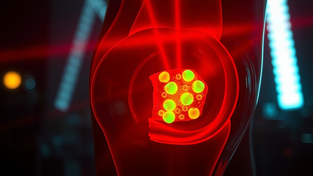
Here's what caught my attention. The study tested four wavelength ranges — 400–405 nm (violet), 500–505 nm (green), 700–710 nm (red), and 1064 nm (near-infrared) — across four energy densities: 3, 15, 30, and 60 J/cm².[1] That's a 16-condition matrix applied to human meniscus-derived stem cells (MeSCs). Most PBM studies pick one wavelength and one dose and call it a day. This one didn't.
The results were unequivocal. Only the 700–710 nm and 1064 nm wavelengths at 3, 15, and 30 J/cm² improved MeSC proliferation and mitochondrial membrane potential. Peak effect: 15 J/cm². Everything else — every violet and green wavelength condition, and every condition at 60 J/cm² — reduced mitochondrial function and proliferative capacity[1].
Let me be direct about what this means. The biphasic dose response in PBM isn't just a theoretical concept. It's a cliff edge. At 60 J/cm², regardless of wavelength, cells performed worse than controls. The wellness market has done more damage to this field than the skeptics ever could, because "more light = more benefit" is exactly wrong.
The Calcium-ROS Signaling Axis#
The mechanistic finding is the real contribution here. Intracellular Ca²⁺ levels and reactive oxygen species (ROS) production increased proportionally with energy density across all wavelengths. But — and this is critical — cytochrome c oxidase (CCO) activity and nitric oxide (NO) concentrations remained unchanged[1].
That last sentence should stop you cold if you've been following PBM research. The dominant hypothesis for two decades has been that PBM works primarily through CCO activation at complex IV of the mitochondrial electron transport chain, displacing NO and boosting ATP synthesis. This study found no evidence for that mechanism in MeSCs.
Instead, when the researchers inhibited the TRPV1 calcium channel, PBM-induced elevations in both Ca²⁺ and ROS were abolished at all wavelengths. Proliferative changes disappeared entirely[1]. TRPV1 isn't just involved — it appears to be the primary mediator.
Mitochondrial Energy Metabolism in Neurodegeneration#
The MeSC findings gain additional weight when read alongside Chen et al.'s work on PBM in APP/PS1 Alzheimer's disease mice, published in Alzheimer's Research & Therapy[2]. Using 808 nm near-infrared light at 20 mW/cm² over six weeks, the team demonstrated that PBM modulated mitochondrial energy metabolism, reduced neuroinflammation, inhibited neuronal apoptosis, and improved cognitive function in AD model mice.
The convergence matters. Two independent studies, different tissue types, different wavelengths — both pointing to mitochondrial energy metabolism as PBM's central target. Chen et al. showed PBM protected mitochondrial dynamics and function, attenuating amyloid-β induced damage[2]. The MeSC study mapped the signaling pathway upstream: TRPV1 → Ca²⁺ influx → controlled ROS → proliferative response[1].
I'm less convinced by the Alzheimer's data as directly translatable to human protocols — it's a mouse model, the APP/PS1 transgenic specifically, and the gap between murine neurodegeneration and human AD remains significant. But the mitochondrial mechanism alignment across studies is hard to dismiss.
MeSC Proliferation Response by Wavelength at 15 J/cm²
COMPARISON TABLE#
| Method | Mechanism | Evidence Level | Cost | Accessibility |
|---|---|---|---|---|
| PBM 700–710 nm LED (15 J/cm²) | TRPV1-Ca²⁺-ROS signaling → mitochondrial function ↑ | Single in vitro study (MeSCs) | Low–Moderate ($50–$300 for calibrated LED panels) | High (home use possible with correct device) |
| PBM 808 nm NIR (20 mW/cm²) | Mitochondrial energy metabolism modulation, anti-neuroinflammation | Preclinical (AD mouse model) | Moderate ($200–$500 for NIR devices) | Moderate (specific irradiance needed) |
| PBM 660 nm (prior MeSC studies) | Chondrogenic differentiation, proliferation | Limited in vitro | Low ($40–$150) | High |
| Platelet-Rich Plasma (PRP) for meniscus | Growth factor delivery, tissue regeneration | Multiple small human RCTs | High ($500–$2,000 per injection) | Low (clinical setting only) |
| Surgical meniscus repair | Mechanical reattachment | Strong clinical evidence | Very High ($5,000–$30,000+) | Low (hospital, recovery time) |
THE PROTOCOL#
Based on the current evidence from this in vitro study, these parameters represent the most promising starting points. I want to be clear: optimal dosing in humans for meniscal or joint applications is not yet established. This protocol extrapolates from cell culture data, and tissue penetration changes everything.
Step 1. Select a light source in the 700–710 nm (red) or 1064 nm (near-infrared) range. Wavelength matters. Irradiance matters. Time matters. Skin matters. Most consumer devices get at least one of these wrong. Verify the manufacturer provides spectral output data — not just a claimed wavelength.
Step 2. Target an energy density of 15 J/cm² at tissue level. This was the peak proliferative response in the study[1]. Calculate exposure time based on your device's irradiance (mW/cm²). Formula: Time (seconds) = Energy density (J/cm²) ÷ Irradiance (W/cm²). For a device outputting 50 mW/cm², that's 300 seconds (5 minutes).
Step 3. Position the light source directly over the target area — for knee/meniscus applications, this means medial or lateral joint line. Maintain consistent distance. Even small changes in distance alter irradiance significantly with LEDs.
Step 4. Apply treatment once daily. The study irradiated cells once and assessed outcomes at multiple time points. For a human protocol, daily application is a reasonable starting frequency, but do not exceed 30 J/cm² per session — the data shows diminishing returns and potential harm above this threshold[1].
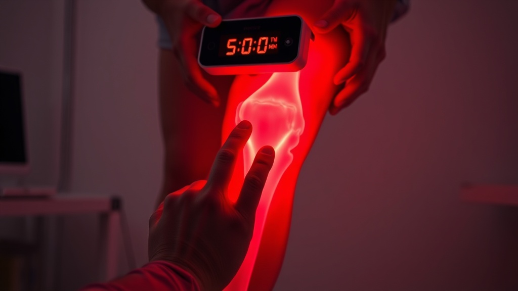
Step 5. Track outcomes. For joint applications, monitor pain levels (VAS scale), range of motion, and functional capacity over a minimum 4–6 week period. Without tracking, you're not experimenting — you're guessing.
Step 6. Avoid stacking with violet (400–405 nm) or green (500–505 nm) wavelengths in the same session. The data explicitly showed these wavelengths reduced mitochondrial function and proliferative capacity at every energy density tested[1]. Multi-wavelength panels that include these ranges may be counterproductive for this application.
Related Video
What is the TRPV1 channel and why does it matter for photobiomodulation?#
TRPV1 (transient receptor potential vanilloid 1) is a calcium-permeable ion channel found in cell membranes throughout the body. This study demonstrated that PBM's effects on stem cell proliferation and mitochondrial function are primarily mediated through TRPV1 activation — when the channel was blocked, all PBM-induced cellular changes were eliminated[1]. This challenges the previous dominant theory that cytochrome c oxidase was the main PBM target.
Why does higher energy density reduce cell function instead of improving it?#
This is the biphasic dose response — sometimes called the Arndt-Schulz curve in PBM literature. At 60 J/cm², intracellular calcium and ROS levels exceeded beneficial thresholds, shifting from signaling molecules to sources of oxidative damage. The data from this study confirmed that the sweet spot for MeSCs sits around 15 J/cm², with 3 and 30 J/cm² still beneficial but less effective[1].
How does this research relate to Alzheimer's disease treatment?#
Chen et al. independently showed that PBM at 808 nm modulated mitochondrial energy metabolism and reduced neuronal damage in Alzheimer's mouse models[2]. While the tissue type and application differ entirely from the MeSC study, both converge on mitochondrial function as PBM's core target. However, all AD data is preclinical — in mouse models — and direct translation to human cognitive protocols requires significant additional research.
What wavelengths should I avoid for joint or cartilage applications?#
Based on this study, violet (400–405 nm) and green (500–505 nm) wavelengths reduced mitochondrial membrane potential and cell proliferation at every energy density tested[1]. If your goal is stem cell proliferation and mitochondrial support for joint tissue, stick to the 700–710 nm or 1064 nm range. I'd honestly avoid multi-spectrum panels that can't isolate wavelengths for this reason.
When will human clinical trials confirm these findings?#
Honestly, we don't know yet. The MeSC study is in vitro — cells in a dish. The AD study is in mice. Clinical translation typically takes years, and the PBM field has historically struggled with standardization of parameters across trials. The specific TRPV1 mechanism identified here may accelerate targeted trial design, but I'd want to see at least a small pilot study in human subjects before changing my protocol with any confidence.
VERDICT#
Score: 7/10
The mechanistic contribution here is genuinely significant. Identifying TRPV1 as the primary mediator — and effectively ruling out CCO in this cell type — is the kind of data point that redirects a field. The 16-condition dose-response matrix is more thorough than most PBM studies I've reviewed, and the results are internally consistent.
But it's still a single in vitro study. No control group issues to complain about — they had proper controls — but cells in culture don't have skin, subcutaneous fat, or systemic circulation competing with photon delivery. The Alzheimer's mouse data from Chen et al. adds mechanistic weight but remains preclinical. I want to see the TRPV1-PBM axis tested in human tissue explants or, better, a small clinical pilot before this moves beyond informed self-experimentation.
The parameters are clear enough to act on if you choose to: 700–710 nm or 1064 nm, 15 J/cm², once daily. That specificity earns points. Most PBM research leaves you guessing. This one doesn't.
References
- 1.Author(s) not listed. Photobiomodulation stimulates mitochondrial function and cell proliferation in meniscus-derived stem cells (MeSCs) via activation of TRPV1 channel. Scientific Reports (2025). ↩
- 2.Chen H, Li N, Liu N, Zhu H, Ma C, Ye Y, Shi X, Luo G, Dong X, Tan T, Wei X, Yin H. Photobiomodulation modulates mitochondrial energy metabolism and ameliorates neurological damage in an APP/PS1 mouse model of Alzheimer's disease. Alzheimer's Research & Therapy (2025). ↩
Sova Reld
Sova writes with focused intensity and low tolerance for vague claims. She came to photobiomodulation through personal experimentation and is irritated by both true believers and reflexive skeptics. Her writing has edge: 'The wellness market has done more damage to this field than the skeptics ever could.' She's extremely precise about parameters — wavelength, irradiance, duration — and will tell you when a study used inadequate dosing without apology.
View all articles →