Single-Cell Atlas Reveals How Breast Tissue Ages at Cellular Level
SNIPPET: A new single-cell spatial atlas of over 3 million cells from 527 human breast tissue samples reveals that aging causes a sweeping decline in cellularity, proliferation, and tissue architecture — with fewer lobules, increased fat, and a shift toward a pro-inflammatory immune microenvironment dominated by M2 macrophages and cytotoxic T cells, fundamentally altering the landscape from which breast cancer emerges.
THE PROTOHUMAN PERSPECTIVE#
The data told me something I didn't expect. Not that breast tissue ages — everything ages — but that it ages this comprehensively, this uniformly. Every compartment declining in concert. Epithelial, stromal, immune. All of it.
For those of us who think about longevity on a systems level, this matters because it reframes breast cancer risk as an architectural problem, not just a mutational one. The tissue itself is remodeling into something more hospitable to disease. Fewer functional lobules, more adipose tissue, a microenvironment that's chronically inflamed and immunologically shifted. This is the kind of finding that should change how we think about cancer prevention — not just screening for mutations, but actively maintaining tissue integrity across decades.
And there's a broader evolutionary angle worth noting. The breast is a hormone-sensitive organ that was never designed, in evolutionary terms, to persist this long past reproductive utility. We're running hardware well beyond its biological warranty. Understanding how that decay unfolds at single-cell resolution is the first step toward doing something about it.
This is not marginal. This is structural.
THE SCIENCE#
What the Atlas Actually Shows#
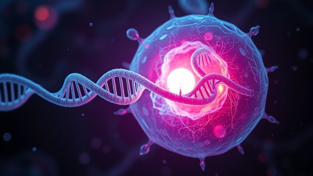
A spatial atlas of the aging human breast is a map of 40 proteins profiled across over 3 million individual cells, sourced from 527 reduction mammoplasty specimens. The research team, publishing in Nature Aging in March 2026, used imaging mass cytometry (IMC) to achieve this — a technique that preserves spatial context, meaning we know not just what the cells are, but where they sit relative to each other [1].
That spatial dimension matters. A lot.
Aged breast tissue was less cellular and less proliferative across all cell types. Epithelial cells, stromal cells, immune cells — all showed reduced numbers and lower proliferative markers with advancing age. The tissue architecture itself restructured: fewer heterotypic epithelial cell–cell interactions, a dramatic reduction in lobular structures, and a corresponding increase in adipose (fat) tissue [1].
The Immune Shift#
Here's where the data gets genuinely interesting. Older breast tissues displayed what the authors describe as a more "inflammatory microenvironment." Specifically, M2 macrophages and granzyme B+ T cells were enriched in aged tissue, while younger tissues were characterized by a relative enrichment of B cells [1].
M2 macrophages are the tissue-remodeling, immunosuppressive phenotype — the ones associated with wound healing but also with tumor-permissive environments. Granzyme B+ T cells are cytotoxic, but their presence alongside M2 macrophages suggests a microenvironment that's simultaneously inflamed and immunosuppressed. That's a dangerous combination.
This aligns with companion work from a mouse mammary gland aging atlas published in Nature Aging in 2024, which found that distinct T cell subsets (Gzmk+, memory CD4+, γδ) and M2-like macrophages expand with age, and that aged epithelial cells exhibit epigenetic changes in metabolic, pro-inflammatory, and cancer-associated genes [2]. The mouse and human data are telling the same story. That convergence is reassuring.
The Senescence Connection#
But here's where it gets complicated. A separate 2026 study in Nature Aging demonstrated that during postpartum mammary gland involution in mice, a p16Ink4a-dependent senescence response is physiologically required for normal tissue remodeling — but can be hijacked by oncogenic events to promote tumorigenesis [3]. Senescent cells enhance tumor cell plasticity via the senescence-associated secretory phenotype (SASP), fostering metastasis.
Let me push back on one thing here: this was done in mouse models, and postpartum involution is a specific biological context. Extrapolating directly to age-related breast tissue remodeling requires caution. The SASP machinery overlaps, but the triggers and timing are different. I'd want to see whether the senescent cell populations in the aging atlas share the same SASP profile as those in involution before connecting these dots too firmly.
The Cancer Bridge#
The third piece of this puzzle comes from work showing that cell populations in human breast cancers are molecularly and biologically distinct with age. Using an Age-Specific Program ENrichment (ASPEN) pipeline, researchers found that in triple-negative breast cancer, aging was associated with increased epithelial–mesenchymal transition in tumor cells and inflammatory responses in cancer-associated fibroblasts [4]. In estrogen receptor-positive cancers, aging correlated with increased ESR1 expression and reduced vascular and immune cell metabolism.
Both younger (<45) and older (>65) patients have worse outcomes than the 55–64 reference group, regardless of subtype — with older patients faring the worst [4]. The atlas data suggests this isn't random bad luck. The aged tissue microenvironment is primed for a different kind of cancer.
The Broader Single-Cell Atlas Landscape#
A complementary human breast cell atlas from Nature Genetics, assembled from more than 800,000 cells across 55 donors, identified 41 cell subclusters and found that age, parity, and germline mutations each affected breast tissue homeostasis differently [5]. Critically, immune cells from BRCA1 or BRCA2 carriers showed gene expression signatures indicative of immune exhaustion — suggesting that immune-escape mechanisms may manifest in non-cancerous tissue well before a tumor develops.
This is the kind of early-detection signal that changes timelines. Not screening for cancer, but screening for the conditions that precede it.
Key Age-Related Changes in Breast Tissue Microenvironment
COMPARISON TABLE#
| Method | Mechanism | Evidence Level | Cost | Accessibility |
|---|---|---|---|---|
| IMC Spatial Atlas (New) | 40-protein spatial profiling via imaging mass cytometry; 3M+ cells, 527 samples | High — large human cohort, published in Nature Aging | Very High (research-grade) | Research institutions only |
| scRNA-seq Atlas | Single-cell RNA sequencing of 800K+ cells across 55 donors | High — published in Nature Genetics | High | Academic/clinical labs |
| Mouse Mammary Aging Atlas | Single-cell transcriptomic + epigenomic profiling in mouse models | Moderate — animal model, requires human validation | Moderate | Research labs |
| Bulk RNA Sequencing | Aggregate gene expression across tissue samples | Moderate — lacks cell-type specificity | Moderate | Widely available |
| Standard Mammography Screening | Radiographic imaging for structural abnormalities | High for detection — misses microenvironment changes | Low–Moderate | Widely accessible |
THE PROTOCOL#
How to act on this data if you're serious about breast tissue health and cancer prevention across decades. Based on current evidence, these are actionable steps — with the caveat that some are extrapolated from preclinical and observational data.
Step 1: Understand your baseline tissue age. If you're undergoing any breast procedure (biopsy, reduction, risk-reduction mastectomy), request that tissue be banked for future single-cell analysis. This isn't standard yet, but tissue banks are expanding, and having a cellular baseline will become increasingly valuable.
Step 2: Target chronic inflammation systemically. The atlas data points to a pro-inflammatory microenvironment as a hallmark of aging breast tissue. Anti-inflammatory interventions with evidence include omega-3 fatty acids (2–4 g/day EPA+DHA), curcumin (500 mg/day bioavailable formulation), and regular aerobic exercise (150+ minutes/week at moderate intensity). These won't reverse tissue aging, but they may slow the inflammatory drift.
Step 3: Monitor immune biomarkers longitudinally. Ask your physician about tracking inflammatory markers — CRP, IL-6, TNF-α — as part of annual panels. The shift from B cell-enriched to M2 macrophage-enriched microenvironments isn't something you can measure from a blood draw yet, but systemic inflammatory markers correlate.
Step 4: Consider senolytic research with extreme caution. The senescence data is provocative — senescent cells contribute to the pro-tumorigenic microenvironment, and senolytics (fisetin, dasatinib + quercetin) have shown preclinical promise. But the involution data shows senescent cells are also required for normal tissue remodeling [3]. Timing matters enormously. Do not self-administer senolytics without clinical guidance.
Step 5: Prioritize mammographic density awareness. Increased adipose replacement with age correlates with changes in mammographic density. Discuss your density category with your radiologist. Dense tissue in older women may warrant supplemental MRI screening, not just standard mammography.
Step 6: If you carry BRCA1/BRCA2 mutations, request immune profiling. The atlas data from Gray et al. showed immune exhaustion signatures in non-cancerous tissue from BRCA carriers [5]. Early immune profiling could identify whether you're already showing these exhaustion patterns years before any malignancy.
Related Video
What is a single-cell spatial atlas of breast tissue?#
It's a high-resolution map of individual cells in breast tissue that captures both what type each cell is (epithelial, immune, stromal) and where it sits relative to other cells. The spatial component is critical — it reveals the architecture and neighborhood interactions that bulk sequencing misses entirely. The atlas published in Nature Aging profiled 40 proteins across over 3 million cells from 527 human samples [1].
How does breast tissue aging differ from aging in other organs?#
The breast is uniquely hormone-sensitive and undergoes repeated remodeling throughout reproductive life. Unlike organs such as the brain, where neuron-specific gene expression remains relatively stable with age [6], breast tissue shows a near-universal decline across every cellular compartment — epithelial, stromal, and immune — with simultaneous architectural restructuring. The scale of coordinated decline was unexpected even to the researchers.
Why do both younger and older breast cancer patients have worse outcomes?#
According to the ASPEN analysis, the tumor microenvironment is molecularly distinct at different ages [4]. Younger patients face more aggressive subtypes like triple-negative breast cancer, while older patients contend with a tissue environment already shifted toward inflammation, reduced immune metabolism, and increased fibroblast senescence. Current clinical trials underrepresent both age extremes, meaning treatments aren't optimized for either group.
What are senolytics and should I take them for breast health?#
Senolytics are compounds that selectively eliminate senescent cells. Preclinical evidence suggests they may reduce the pro-tumorigenic SASP in mammary tissue [3]. However — and this is critical — senescent cells also serve essential physiological roles in tissue remodeling. Removing them at the wrong time could disrupt normal processes. Optimal dosing and timing in humans is not yet established. This is not a supplement to take casually.
How can I monitor my breast tissue aging without a biopsy?#
Currently, direct cellular-level monitoring requires tissue sampling. However, systemic inflammatory markers (CRP, IL-6), mammographic density assessments, and emerging liquid biopsy technologies offer indirect proxies. The field is moving toward non-invasive approaches, but we're not there yet. Honestly, this is a gap in clinical tools that needs closing.
VERDICT#
Score: 8.5/10
This atlas moved me. The scale — 3 million cells, 527 samples, 40 proteins with spatial resolution — is the most detailed map of breast tissue aging in humans to date. The convergence with mouse data strengthens the findings. The immune shift toward M2 macrophages and cytotoxic T cells in aged tissue is the kind of mechanistic insight that could reshape how we think about cancer prevention, not just detection.
Where I'm less convinced: the clinical actionability is still limited. We know the landscape changes. We don't yet have validated interventions that target those specific changes in living tissue. The senescence story is also double-edged — the same cells that create risk also serve essential functions, and the involution data was done in mice.
But as a foundation for the next decade of breast cancer prevention research, this is the reference point. Everything from here builds on what this atlas reveals.
References
- 1.Author(s) not listed. Single-cell spatial atlas of the aging human breast. Nature Aging (2026). ↩
- 2.Author(s) not listed. Comprehensive single-cell aging atlas of healthy mammary tissues reveals shared epigenomic and transcriptomic signatures of aging and cancer. Nature Aging (2024). ↩
- 3.Author(s) not listed. Induction of senescence during postpartum mammary gland involution supports tissue remodeling and promotes postpartum tumorigenesis. Nature Aging (2026). ↩
- 4.Author(s) not listed. Cell populations in human breast cancers are molecularly and biologically distinct with age. Nature Aging (2025). ↩
- 5.Gray GK et al.. A single-cell atlas enables mapping of homeostatic cellular shifts in the adult human breast. Nature Genetics (2024). ↩
- 6.Author(s) not listed. Single-cell transcriptomic and genomic changes in the ageing human brain. Nature (2025). ↩
Orren Falk
Orren writes with the seriousness of someone who thinks about their own mortality every day and has made peace with it. He takes the long view, which means he's less excited than others about marginal gains and more focused on whether something moves the needle on a decade-level timescale. He'll admit when a study impresses him: 'This one actually moved me.' He uses 'the data' as a character in his writing — it speaks, it tells him things, it sometimes disappoints him.
View all articles →